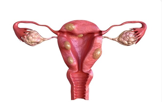
Fibroids
What is a fibroid?
Fibroids are non-cancerous growths that grow or develop in or near the uterus.
The growths are formed of muscle and fibrous tissue, and their size varies. They are sometimes referred to as uterine myomas or leiomyomas.

Types of Fibroids
Fibroids are classified into three types:
- Subserosal fibroids: These are the most prevalent types of fibroids. They have the ability to push outside of the uterus and into the pelvis. Subserosal fibroids can become large and have a stalk that attaches to the uterus at times (pedunculated fibroid).
- Intramural fibroids: These fibroids form in the uterine muscle wall.
- Submucosal fibroids: These are uncommon fibroids. They have the ability to develop into the open space inside the uterus and may also have a stalk.
What are the causes of Fibroids?
The cause of uterine fibroids is unknown, while research suggests that there may be a genetic link. There is no specific external exposure that can cause a woman to develop fibroids.
What are the Signs and Symptoms of Fibroids in the Uterus?
The majority of women with fibroids will have no symptoms at all. Large or several fibroids, on the other hand, might cause the following symptoms:
- Heavy or prolonged periods
- Bleeding between menstrual periods
- Pelvic discomfort and pressure
- Frequent urination
- Pain in the lower back region
- Pain during intercourse
- Difficulties to get pregnant
Diagnostic Methods for Fibroids
Fibroids are most commonly discovered during a physical examination. During an abdominal or pelvic exam, your doctor may feel a solid, irregular (often painless) mass.
- Ultrasound: The most widely utilised scan for fibroids is ultrasound. It diagnoses fibroids using sound waves at frequencies. To scan the uterus and ovaries, a doctor or technician puts an ultrasound probe on the abdomen or into the vagina. It is quick, easy to use, and generally accurate. For other disorders, such as adenomyosis, alternative testing, such as MRI, may be more effective.
- MRI: This imaging technique creates images using magnets and radio waves. It provides your physician about the size, quantity, and location of the fibroids. Doctors can also tell the difference between fibroids and adenomyosis, which is commonly misdiagnosed. MRI is used to confirm a diagnosis and to help us decide which treatments are best for you. In addition, MRI may be a superior option for similar disorders such as adenomyosis.
- Hysterosalpingogram (HSG): A HSG is often used by doctors for women who are having difficulty conceiving. It examines the uterus (uterine cavity) and fallopian tubes. After inserting a catheter (small tube) into the uterus, the doctor slowly injects a specific contrast dye and obtains X-rays.
-
Hysterosonogram: A hysterosonogram allows doctors to see within the uterus. They inject water into the uterus after inserting a tiny catheter and collecting a series of ultrasound photos. The test can detect uterine polyps or intracavitary fibroids, which can cause severe bleeding.
-
Hysteroscopy: A doctor uses a long, thin tool with a camera and light to examine possible problems inside the uterus. The tool is passed via the vagina and cervix into the uterus by the doctor. There is no need for an incision. Using this method, the doctor can examine for fibroids or endometrial polyps within the uterine cavity. During this treatment, your doctor may also remove some forms of fibroids.
What are the risks of uterine fibroids?
It is unusual for fibroids to have serious health implications. Women, on the other hand, can experience severe bleeding, which can result in hazardous anaemia, or a lack of red blood cells.
Large fibroids can occasionally push on the bladder and the tube (ureter) that transports urine from the kidney to the bladder. This pressure can harm the kidneys. Infertility and frequent pregnancy loss are two more problems that can arise.
Treatment for Fibroids
Medical Treatment:
Anti-inflammatory pain relievers such as ibuprofen or naproxen may reduce fibroids-related menstrual flow and give pain relief. This is the most conservative treatment option and is advised for women who experience intermittent pelvic pain or discomfort as a result of fibroids.
Hormonal Therapy:
- Agonists of gonadotropin-releasing hormone (GnRH agonists). This treatment reduces your oestrogen levels and causes a brief “medical menopause.” To shrink the fibroid, GnRH agonists are employed (s). They can also be used to delay your menstruation before surgery or to increase your blood count.
- Oral contraceptive tablets (or a patch or vaginal ring) can help minimise fibroids-related bleeding.
- Progesterone-containing medications, such as tablets, implants, injections, or intrauterine devices (IUDs), may also be used to control bleeding.
Surgical Treatment:
Conservative surgical treatment. Myomectomy is a treatment that removes fibroids while leaving the uterus intact. This method is suggested for women who want to keep their fertility. There are three basic procedures of myomectomy:
Open Myomectomy: This is the most common type of myomectomy. The operation is done through an abdominal incision and entails some hazards, such as haemorrhage and scar tissue formation at the incision site, as well as a prolonged recovery time. Depending on the size and quantity of fibroids, this method may be required.
Depending on the size and quantity of fibroids, this method may be required.
Robotic Myomectomy:
Myomectomy can be performed laparoscopically or robotically. A laparoscope and small “keyhole” abdominal incisions are used in this outpatient surgery. This less invasive method frequently leads to less bleeding and a quicker recovery, although it is not appropriate for all people. Most patients go home the same day as their operation and recover in a few weeks.
Hysteroscopic Myomectomy:
Myomectomy through hysteroscopic surgery. During this outpatient treatment, your doctor will cut off visible portions of the fibroid tumours with a camera introduced through the vagina. This procedure is exclusively used to treat fibroids that have developed within the uterine cavity.
Uterine Artery Embolization (UAE): Commonly known as uterine fibroid embolization, is a relatively new procedure. By cutting off the blood flow to fibroids, this minimally invasive treatment reduces them. UAE is performed by an interventional radiologist with the assistance of X-rays.
Another newer procedure is radiofrequency ablation of fibroids, in which heat is administered to the fibroids under laparoscopic and ultrasound guidance to shrink and soften them. The potential consequences on fertility are still unknown.
Hysterectomy:
Depending on the size of your uterus, the location of the fibroids, and your medical history, the treatment can be performed vaginally or abdominally through a major incision, laparoscopically or robotically.
Because a hysterectomy is a significant procedure, it is only indicated to treat fibroid cases in women who do not want to keep their fertility. Because it removes the potential of recurrence, it is the most effective method of fibroid treatment.
KK speciality clinic and hospital is one of the best hospital in Western suburbs of Mumbai for all types of laproscopic surgery and treatment for fibroids.
